Home › Without Label › Peritoneal Mesothelioma Ct Scan - Bidirectional Chemotherapy Allowing Surgery And Hipec For Malignant Peritoneal Mesothelioma - peritoneal inclusion cyst is a less common benign primary peritoneal tumor.
Peritoneal Mesothelioma Ct Scan - Bidirectional Chemotherapy Allowing Surgery And Hipec For Malignant Peritoneal Mesothelioma - peritoneal inclusion cyst is a less common benign primary peritoneal tumor.
Peritoneal Mesothelioma Ct Scan - Bidirectional Chemotherapy Allowing Surgery And Hipec For Malignant Peritoneal Mesothelioma - peritoneal inclusion cyst is a less common benign primary peritoneal tumor.. The clinical presentations and imaging findings are nonspecific and resemble various diseases, including peritoneal metastasis. ct scan for mesothelioma a ct scan generates detailed three dimensional images of specific regions of the body. Because adequate cytoreduction is necessary to achieve prolonged survival, ct scans became an accurate prognostic radiologic test for patient selection for comprehensive treatment. Malignant peritoneal mesothelioma (mpm) is a rare but fatal tumor. It is usually associated with asbestos exposure and regarded as universally fatal.
Your general practitioner (gp) will assess your symptoms. The second patient was also examined with mri. A ct scan may be performed in order to diagnose peritoneal mesothelioma. Radiology imaging sometimes will not pick up evidence of diffuse cancerous growths on the mesothelium, but will readily produce a shadow created by a single tumor that may occur more readily with peritoneal mesothelioma. You may have an abdominal ultrasound scan to check for peritoneal mesothelioma.
The Radiology Assistant Peritoneal Pathology from radiologyassistant.nl The data gathered by the ct scan is used to work out one of the simplest ways of acquiring tissue for testing (see biopsy beneath). ct scans are often used to help look for mesothelioma and to find the exact location of the cancer. People with peritoneal mesothelioma may have fluid in the abdomen (called peritoneal effusion), causing swelling and pain. Using an ultrasound scan to guide the doctor, the fluid is drained via a needle inserted through the chest wall into the pleural cavity or into the abdomen. Researchers at the university of chicago want to find out if the new experimental mri and ultrasound imaging techniques do a better job of detecting these cancers. peritoneal mesothelioma is a rare cancer of the abdominal lining with about 600 cases per year in the united states. Your gp will conduct a physical examination and order tests. If determined that you do have peritoneal mesothelioma, it is very important to get an evaluation at a healthcare facility with expertise in the condition since it is so rare.
Find out how a ct scan can help to diagnose mesothelioma and what happens during the test.
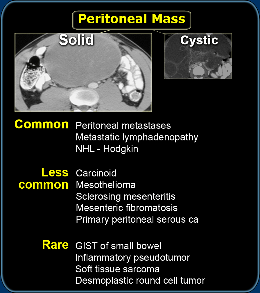
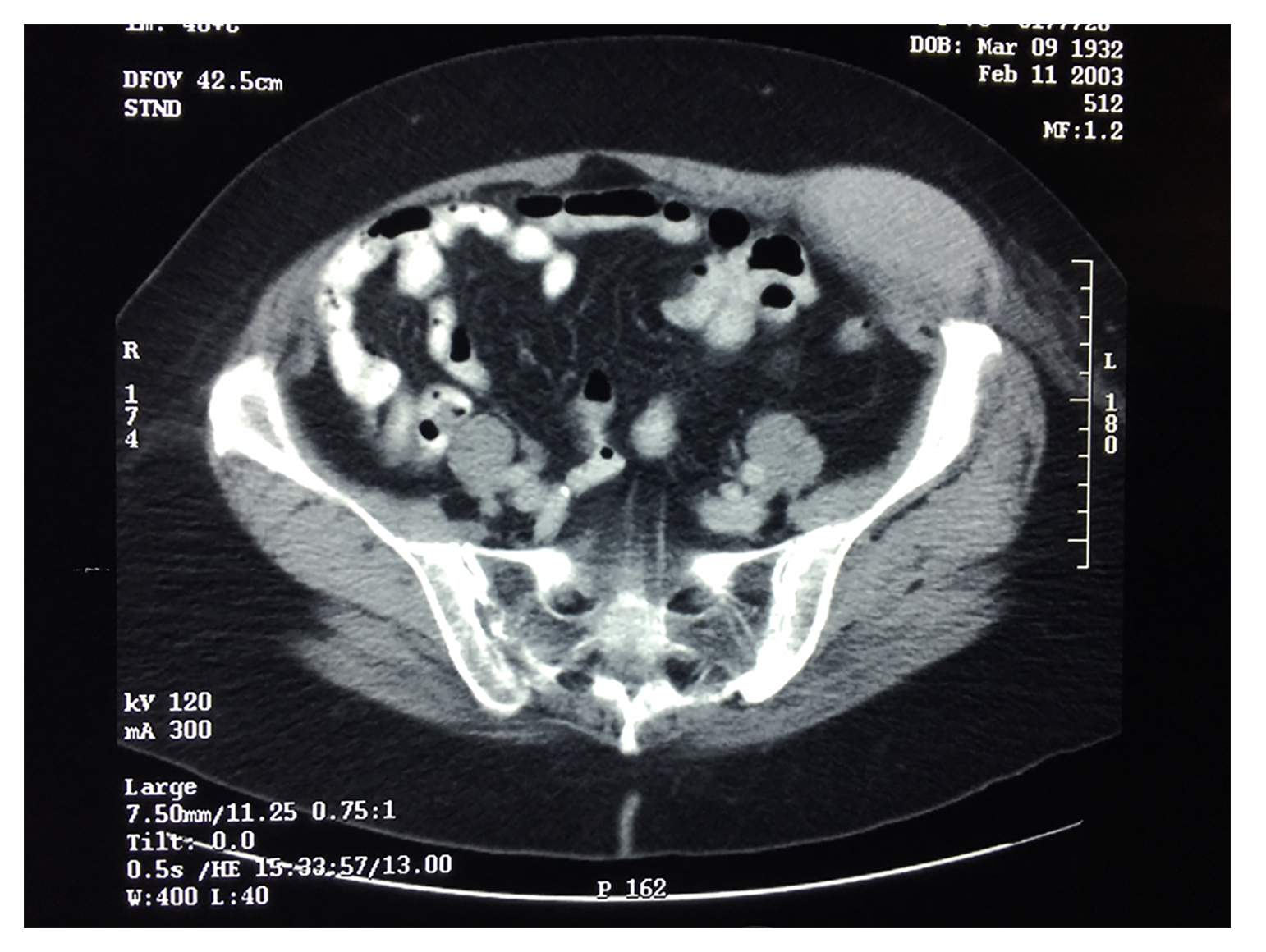
According to the national cancer institute, the peritoneum is the layer of tissue that lines the entire abdominal wall and essentially covers all of the. Report of 11 new cases and review of the literature. Find out how a ct scan can help to diagnose mesothelioma and what happens during the test. Clinical symptoms and findings may be confusing and diagnosis can be easily overlooked especially in cases where there is no previous asbestos exposure. Imaging scans identify the location and size of mesothelioma tumors. The reason for this is that doctors can easily confuse mesothelioma on a ct scan or mri with an abundance of gas and misdiagnose the disease as a result. A ct scan may suggest the presence of cancer in the abdominal cavity, but cannot confirm the diagnosis of mpm. peritoneal mesothelioma 1 and 2 axial contrast enhanced ct scans show abundant ascites a with micronodular invasio mesothelioma disease treatment pleural effusion pericardial effusion cancer / malignant mesothelioma is a rare, aggressive cancer that attacks the lining of internal organs. The standard time delay between administration of contrast and ct imaging acquisition is. A ct scan can show abnormal swellings in the lining of your lungs (pleura) or abdomen (peritoneum). A biopsy is always required for confirmation. For cancers, such as mesothelioma, that spread to the lining of the stomach, detecting the cancer is very difficult with ct or mri scans. An analysis of the preoperative ct scans of 30 patients with peritoneal mesothelioma treated with cytoreductive surgery and perioperative intraperitoneal chemotherapy at a single.
When asbestos fibers become lodged in the lungs and trachea, they can make their way into the abdomen. Your general practitioner (gp) will assess your symptoms. Researchers at the university of chicago want to find out if the new experimental mri and ultrasound imaging techniques do a better job of detecting these cancers. A ct scan is approximately 90% sensitive for detecting malignant pleural mesothelioma. It is also known as benign multicystic mesothelioma.
Scielo Brasil Benign Multicystic Peritoneal Mesothelioma Literature Review And Update Benign Multicystic Peritoneal Mesothelioma Literature Review And Update from minio.scielo.br Imaging studies are helpful in evaluating patients who have symptoms suggestive of peritoneal carcinomatosis. In mpm, the ct scan identifies a solid, heterogeneous mass with irregular margins. If you have signs and symptoms that might indicate mesothelioma, your doctor will conduct a physical exam to check for any lumps or other unusual signs. For instance, they can show if the cancer has spread to other. This case report describes the imaging findings of malignant peritoneal mesothelioma and the additional value of pet/ct. Pet/ct scan problem of pleural effusion therapy for mesothelioma sufferers. This information is an important part of planning mesothelioma treatments. Suggest mesothelioma include an abnormal thickening of the pleura, calcium deposits on the pleura, fluid in the space between the lungs and the chest wall, or changes in the lungs themselves as a result of asbestos exposure.
A ct scan may suggest the presence of cancer in the abdominal cavity, but cannot confirm the diagnosis of mpm.
mesothelioma tests commonly include imaging scans, such as mri and ct scans, and biopsies such as pleural aspiration or thoracoscopy. The standard time delay between administration of contrast and ct imaging acquisition is. The journal of thoracic disease indicates that not only does mesothelioma show up on a ct scan but it is the preferred diagnostic tool of choice for advanced stage mesothelioma cases. It might probably additionally present if the mesothelioma has unfolded to different organs. You may have several different tests to help diagnose peritoneal mesothelioma. Malignant peritoneal mesothelioma can be difficult to diagnose. Your gp will conduct a physical examination and order tests. It is usually associated with asbestos exposure and regarded as universally fatal. peritoneal mesothelioma is a rare cancer of the abdominal lining with about 600 cases per year in the united states. The ct scan supplies correct details about the situation and thickness of the tumour(s) within the chest or stomach. A ct scan can show abnormal swellings in the lining of your lungs (pleura) or abdomen (peritoneum). Worrisome lesions seen on ct imaging would prompt a biopsy attempt to confirm the diagnosis. However, they can help the doctor understand the extent and stage of disease.
This information is an important part of planning mesothelioma treatments. The second patient was also examined with mri. peritoneal mesothelioma is a rare form of cancer that develops in the lining (mesothelium) of the abdomen (peritoneum), covering the surface of the omentum and visceral organs. ct scans are often used to help look for mesothelioma and to find the exact location of the cancer. A ct scan may suggest the presence of cancer in the abdominal cavity, but cannot confirm the diagnosis of mpm.
Unusual Radiologic Presentations Of Malignant Peritoneal Mesothelioma from f6publishing.blob.core.windows.net peritoneal mesothelioma 1 and 2 axial contrast enhanced ct scans show abundant ascites a with micronodular invasio mesothelioma disease treatment pleural effusion pericardial effusion cancer / malignant mesothelioma is a rare, aggressive cancer that attacks the lining of internal organs. A ct scan may suggest the presence of cancer in the abdominal cavity, but cannot confirm the diagnosis of mpm. ct scans are often used to help look for mesothelioma and to find the exact location of the cancer. The ct findings were evaluated for the morphologic appearance of. Standard imaging tests, including ultrasonography and helical computed tomography (ct) scans, are notably insensitive for the detection of peritoneal tumors. The second patient was also examined with mri. Malignant peritoneal mesothelioma (mpm) is a rare but highly aggressive malignancy arising from the serosal linings of the peritoneal cavity. Imaging is important in diagnosing mesothelioma.it will provide information such as the extent of disease in the original organ and also show if the cancer has spread to.
Standard imaging tests, including ultrasonography and helical computed tomography (ct) scans, are notably insensitive for the detection of peritoneal tumors.
They can also help determine the stage (extent) of the cancer. peritoneal mesothelioma 1 and 2 axial contrast enhanced ct scans show abundant ascites a with micronodular invasio mesothelioma disease treatment pleural effusion pericardial effusion cancer / malignant mesothelioma is a rare, aggressive cancer that attacks the lining of internal organs. It is also known as benign multicystic mesothelioma. Because adequate cytoreduction is necessary to achieve prolonged survival, ct scans became an accurate prognostic radiologic test for patient selection for comprehensive treatment. Imaging scans identify the location and size of mesothelioma tumors. It is seen in women with prior gynaecological surgery or infection. A ct scan can show abnormal swellings in the lining of your lungs (pleura) or abdomen (peritoneum). ct findings and serum ca 125 levels in malignant peritoneal mesothelioma: Malignant peritoneal mesothelioma (mpm) is a rare but fatal tumor. ct scan for mesothelioma a ct scan generates detailed three dimensional images of specific regions of the body. You may have an abdominal ultrasound scan to check for peritoneal mesothelioma. Doctors also use mesothelioma blood tests to measure treatment response. Both patients had ct scans of the abdomen.


Post a Comment
Post a Comment